Learning
Complete your training modules to enhance your knowledge

Precision in Practice: Mastering Topcon Diagnostic Technology
Learn the core capabilities of Topcon diagnostic technology and how to apply one-touch capture workflows in daily practice.
From Scan to Strategy: Clinical Applications with Topcon Imaging
Connect imaging outputs to practical treatment strategy with examples built around Topcon diagnostic use cases.
Elevating Eye Care: Advanced Workflows
Explore practical ways to streamline advanced workflows across measurement, imaging, and communication in eye care.
Advanced OCT Interpretation
Deep dive into OCT imaging and clinical interpretation.
Fundus Photography Basics
Essential techniques for quality fundus photography.
Tonometry Best Practices
Accurate IOP measurement and device calibration.
Keratometry and Refraction
Streamlining refractive workflows with modern devices.
Pachymetry and Corneal Analysis
Corneal thickness and morphology in clinical practice.
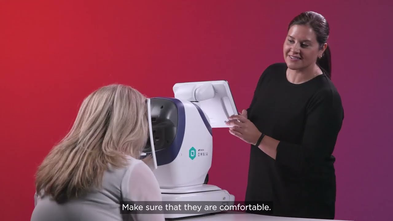
OMNIA Workflow Advanced
Advanced one-touch capture and reporting workflows.
Aladdin Device Overview
Overview of Aladdin system capabilities and setup.
Retinal Disease Recognition
Identifying common retinal findings in imaging.
Glaucoma Screening Protocols
Structured approaches to glaucoma screening and follow-up.
Dry Eye Assessment Tools
Using imaging and metrics in dry eye evaluation.
Pediatric Eye Examination
Adapting workflows for pediatric patients.
Diabetes and Retinal Imaging
Diabetic retinopathy screening and documentation.
Macular OCT Patterns
Recognizing macular pathology on OCT.
Visual Field and Imaging Correlation
Linking structural and functional findings.
EHR and Imaging Integration
Connecting devices and EHR for efficient documentation.
Practice Marketing with Technology
Using diagnostic technology in practice marketing.
Cataract Pre-Op Measurements
Biometry and IOL selection workflows.
Anterior Segment Imaging
Slit-lamp and anterior segment documentation.
Contact Lens Fitting and Imaging
Imaging support for contact lens fitting.
Low Vision Assessment Tools
Tools and workflows for low vision assessment.
Telehealth and Remote Imaging
Remote review and telehealth integration.
Quality Assurance in Imaging
QA and calibration for consistent image quality.
Coding and Reimbursement for Diagnostics
Coding and documentation for diagnostic testing.
Multi-Site Practice Workflows
Scaling workflows across multiple locations.
New Staff Onboarding and Training
Training new staff on devices and workflows.
Retinal Laser and Imaging
Imaging support for laser and treatment planning.
Uveitis and Inflammatory Imaging
Documenting and monitoring inflammatory conditions.
Neuro-Ophthalmology Imaging
Imaging in neuro-ophthalmology assessment.
Ocular Surface Disease Series
Comprehensive ocular surface evaluation.
Myopia Management and Imaging
Imaging in myopia management programs.
Premium IOL Workup and Counseling
Imaging and metrics for premium IOL selection.
Glaucoma Imaging and Progression
Imaging in glaucoma diagnosis and progression.
Retinal Vascular Disease
Imaging and assessment of vascular conditions.
Posterior Segment Trauma
Documentation and follow-up of posterior trauma.
Inherited Retinal Disease
Imaging patterns in inherited retinal conditions.
AMD Management and Imaging
Imaging in age-related macular degeneration care.
VR and AR in Eye Care
Emerging tech and patient education.
Patient Communication and Imaging
Using images to communicate with patients.
Regulatory and Compliance Overview
Device and documentation compliance basics.
Disinfection and Device Care
Cleaning and maintenance for ophthalmic devices.
Inventory and Supply for Diagnostics
Managing supplies and consumables.
Clinical Research and Imaging
Using imaging in clinical research protocols.
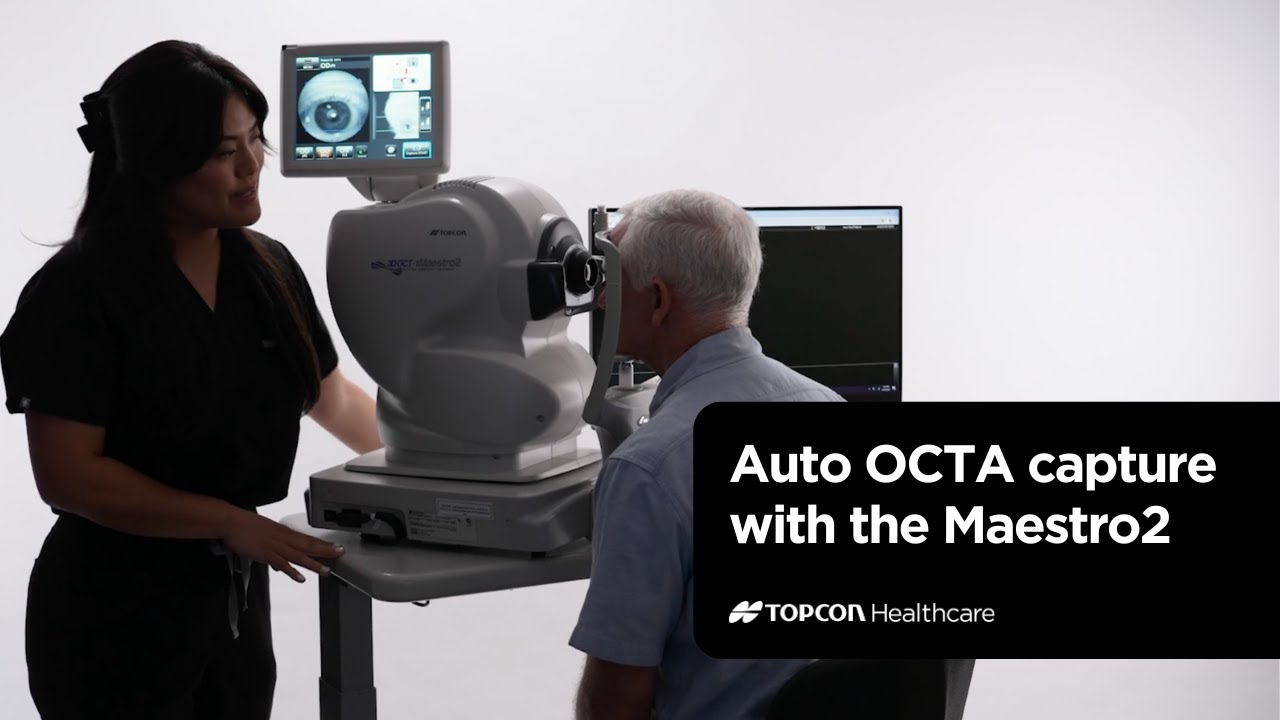
OCT-A Interpretation
OCT angiography fundamentals and interpretation.
Widefield Imaging Techniques
Widefield and ultra-widefield imaging workflows.
B-Scan and Posterior Segment
B-scan ultrasound in posterior segment assessment.
Anterior Segment OCT
AS-OCT in cornea and anterior segment.
Corneal Topography and Mapping
Topography and elevation in clinical practice.
Endothelial Cell Analysis
Specular microscopy and endothelial assessment.